Tuberculosis: Symptoms and Treatment

1 Comment
What is Tuberculosis? Tuberculosis including zoonotic disease, because the disease can be transmitted from animals to humans, for example cows. The most important cause of tuberculosis that causes health problems in many countries around the world is Mycobacterium tuberculosis.

Tuberculosis

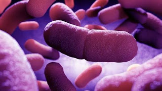
Morphology germs of tuberculosis, Mycobacterium is a rod-shaped bacteria that are often pleomorphism, measuring about 1-4 microns x 0.2-0.5 microns. Germs that the staining including Gram-positive, is resistant to acid and aerobic.

Transmission of tuberculosis, TB germs are transmitted from a human being to another through respiratory air. In addition intestinal tuberculosis can occur if infected with TB germs through cow's milk for tuberculosis. Germs can also be spread through skin inoculation. After entry into the body, the bacteria will spread to the lungs, and then along the blood and lymph spread to many other visceral organs.

Clinical symptoms of tuberculosis, clinical symptoms that occur depending on the type of organ infected with these germs. Infection of the lungs (pulmonary tuberculosis) will cause symptoms of chronic cough, and sometimes bloody sputum (hemoptysis). However often the patient does not show clinical symptoms or real complaint for many years (asymptomatic).

Common symptoms of Tuberculosis are anorexia and weight loss, the body feels tired and lethargic, fever and chills often. In TB skin disorders such as ulcer or papule that develops into a dark-colored pustules.
Miller TB is tuberculosis that attacks various organs ubuh, found in infants or elderly patients with body resistance is low.

Treatment of tuberculosis, treatment of tuberculosis should use more combinations and single drugs, and given the long term continuous, uninterrupted in the middle of the treatment schedule. One way combination treatments for tuberculosis are as follows:
Week 1-4: Rifampicin 450 mg + 400 mg + INH pirasinamid 1,500 mg (given every day). Sunday 5-17: INH Rifampicin 600 mg + 700 mg (given 2 times a week).
Symptomatic treatment is given to subside cough, stop bleeding and other disorders, whereas supportive treatment is given to improving the health and patient endurance.

Prevention of tuberculosis, vaccination with BCG (Bacille Calmette-Guerin came and Mycobacterium bovis) should be administered regularly to the public. Public health education related to prevention of tuberculosis, always drink cow's milk that has been cooked or pasteurized and the careful work in the laboratory at the time dealing with experimental animals, especially primates.

Dengue Fever: Symptoms, Preventions

Add Comment
What's dengue fever? Dengue fever and dengue hemorrhagic fever is a viral disease that is widespread throughout the world, especially in the tropics. Sufferers are mainly children under the age of 15 years, but now many adults this viral disease. The main source of transmission is human and primates, being of vector is the Aedes mosquito.
dengue


Dengue virus, The virus that causes dengue fever is a virus dengue flavivirus genus which includes Arbovirus (Arthropod Borne Virus) group B virus virions have a size of 40 nm. Serologically there are four types of dengue virus, the dengue virus type 1, type 2, type 3 and type 4 virus can breed in a wide variety of tissue culture, eg mammalian cells BHK (Baby Hamster Kidney Cell) and arthropods cells, for example Aedes albopictus cell.

Transmission of dengue fever
Dengue fever is endemic in both urban (urban) and rural (rural). In urban areas the main transmitting vector is Aedes aegypti in urban areas whereas Aedes albopictus. But it often happens that both species of mosquitoes are found together in one area, for example in the areas that are semi-urban. Primates in the forests can act as a source of transmission of infection.

The clinical symptoms of dengue virus infection, The incubation period of dengue fever in humans lasts about 4-5 days. The early symptoms of dengue fever that lasts 1-5 days non-specific, such as mild fever, headache and malaise. Sudden fever occurs within 2-7 days down to normal temperature. Other clinical symptoms that can occur in the form of anorexia, back pain, bone and joint pain.

Dengue hemorrhagic fever (DHF). Manifestations of dengue in the form of bleeding usually occurs on the second day of fever. Bleeding into the skin easily seen if the test tourniquet. Bleeding is also apt to occur at the time of the venipuncture. Of bleeding may be petechiae, purpura, epistaxis and sometimes bleeding gums, hematemesis and melena. Complaints of pain in the abdomen showed the occurrence of gastrointestinal bleeding and shock. At the onset of fever, the patient showed hepatomegaly is usually followed by the shock that occurs on days 3 since pain sufferers.

On examination of the patient's blood platelet blood picture shows a low (less than 100,000 per ml) hematocrit of more than 20% in the second examination, and Sahli hemoglobin levels over 20%.
Serology, for example, complement fixation test, agglutination inhibition test and neutralization assay support the establishment of a diagnosis of dengue fever and dengue hemorrhagic fever.
Isolation of dengue virus and the patient's blood is done by culturing in tissue culture, for example BHK cell culture.

Handling of dengue and DHF
There are no antiviral drugs to combat the dengue virus. DHF patients overcome bleeding and of shock, improved patient endurance, and symptomatic treatment given to relieve patient complaints.

Prevention of transmission of dengue virus
Eradicate the Aedes mosquito is the best way to prevent the spread of dengue virus. Eradication of adult mosquitoes and mosquito larvae must be done together with the eradication of mosquito breeding. Additionally repellen can be used to prevent mosquito bites.

Melasma: Symptoms and Causes

1 Comment
What is that melasma? Chloasma or melasma is a skin disorder in the form of patches with the formation of the dye bark excessively, colored black and brown, are generally symmetrical, uneven, such as discoloration of the skin, often found on the face, the cheeks, forehead, chin, upper lip , and can extend up to the neck.

melasma

Generally occurs in pregnant women or users of oral contraceptives (eg, birth control pills). Melasma is excessive melanin formation caused by exposure to ultraviolet and sunlight.

The cause of melasma, Trends genetics, hormonal influences, and ultraviolet radiation is the most important cause of melasma. More than 30% had a family history of melasma patients who also suffer from melasma. Oral contraceptive pills that contain estrogen-progesterone therapy for prostate cancer can also cause melasma. Medical therapy of allergies and certain cosmetics reported cause melasma, though rarely.
Melasma is rare before the age of puberty. Commonly experienced by adults aged 30-44 years (productive age) and is more common in women. In Indonesia, a comparison of this case to the women and men is 24: 1.

Clinical symptoms of melasma disease begins with patches of black and brown on the cheek, and then extended to the entire face. Skin disorders melasma, hyperpigmentation Plaques with no firm boundaries. Injuries and disorders are usually symmetrical, especially when on the cheek, spread to resemble a mask.
Location disease melasma may occur on the entire face, but mostly on the cheeks, forehead, chin, under the nose, below the lower jaw.

Melasma examination, to determine the type of melasma, Wood lamp examination (with a wavelength of 340-400 nm). There is thickening of the outermost layer of skin; the formation of excessive keratin, but relatively mild. In the basal cells and suprabasal found to deposit melanin. Sometimes melanin is found in all layers of keratinocytes in the epidermis. Melasma Area and Severity Index (MASI) commonly used doctors to assess the condition of patients with melasma. Consider the results of tests to evaluate thyroid function. Not required laboratory tests.

Handling melasma, medications antipigmentasi (depigmenting agents), adjusted for the skin condition of each patient. Hydrogen peroxide and topical corticosteroids. Vitamin C high dose. Vitamin C is thought to inhibit the oxidation dopakuinon. Glutathione. Glutathione is a tripeptide which has a -SH group, may join the cupric ions (Cu) and the enzyme tyrosinase that disrupted melanin formation. The use of bleach (bleaching), which is able to inhibit the activity of the enzyme tyrosinase in the melanocytes, resulting in the formation of melanin is reduced. Retinoids, serves to regulate cell growth and proliferation. For example: topical tretinoin. The use of retinoic acid would affect the pigments in the keratinocytes, also interfere transferpigmen. The use of tretinoin efektifsebagai single therapy. Improvements achieved in a minimum of 6 months.
Laser surgery, using a Q-switched ruby ​​laser and argon laser. Chemical exfoliation, performed by a dermatologist or beautician. Antibiotics, Mass lnya: azelaic acid cream (20%).

Topical therapy approved by the US Food and Drug Administration (FDA) for the treatment of melasma is Triple Combination Cream, containing hydroquinone (4%), tretinoin (0.05%), and fluocinolone acetonide (0.01%). Triple Combination cream is safe and effective when used alternately or continuously for 24 weeks.
Combination treatment with the triple combination cream followed by a laser (1064-nm Q-switched Nd: YAG) proved to be effective, not followed by serious side effects. Topical therapy should be initiated at least eight weeks prior to laser therapy to achieve optimal results.
In topical therapy with a concentration of 4% or less, only a few patients experienced severe side effects.
Generally, the longer the treatment of melasma about 4-6 weeks. Although many treatment products in the form of creams, ointments, capsules, extracts, and others that aims to overcome melasma, be sure to use it with a doctor's supervision. This is to avoid complications or irritations that actually worsen the situation. Better to come immediately to the doctor or dermatologist to get treatment and good handling and measurable.

Melasma can be prevented by: Avoiding the sun, wearing a hat, umbrella, sunscreen and Stopping the use of contraceptives.
The hormone estrogen, progesterone, and MSH (melanocyte-stimulating hormone) can stimulate the occurrence of melasma. Ultraviolet light can stimulate pigment formation process. Melanin (melanogenesis).

Systemic drugs such as chloroquine, chlorpromazine, cytostatic, and minocycline can stimulate melanogenesis. Melasma can occur in men and women who are not pregnant and not using the contraceptive pill.

Malaria: Causes, Symptoms, Treatments

Add Comment
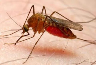
What's Malaria? Malaria is an infectious disease that remains a global health problem. More than one billion people living in malaria-endemic areas, every year millions of people died, mostly children under five years old.

malaria

The cause of malaria, Malaria in humans is caused by four species of Plasmodium, namely Plasmodium vivax, Pl. falciparum, Pl. malariae and Pl. ovale. Types of malaria caused by four type is of the plasmodium malaria cause different patterns of fever or symptoms of clinical symptoms thereof.
Vivax malaria causing Plasmodium vivax, also called benign tertian malaria (benign), being Pl. falciparum causing malaria falciparum malaria tertian or malignant (malignant). Additionally Pl. falciparum malaria also poses perniciosa and Blackwater Fever. PI. malariae cause malaria malariae, and PI. Ovale cause ovale malaria.

Morphological Plasmodium
In the cells of the liver parenchyma, plasmodium is obtained in the form schizonts preeritrositik that for every species of Plasmodium different sizes and number of merozoites in it. Preeritrositik schizonts shape has never been found in Pl. malariae.

Trofozoit. Trofozoit shape plasmadium distinguished young trofozoit and trofozoit further. In Pl. vivax, trophozoite ring-shaped and contain spots basophils, alu-shaped trofozoit amuboid containing spots Schuffner (Schuffner dots). Infected erythrocytes appear enlarged. In trofozoit Further, it appears the pigment parasites, and often more than one parasite inside the erythrocytes (double infection).
In Pl. falciparum, a young ring-shaped trophozoites looked nucleated and cytoplasmic portion is located on the edge and enitrosit (accole or applique form). Often encountered more than one parasite infection with double chromatin spots. Trofozoit further on this species contains spots Maurer (Maurer dots).
PI. malariae has a young trophozoite ring-shaped, with infected erythrocytes are not enlarged. Further trofozoit ribbon-shaped (band-form) and not found Schuffner spots.
Morphology trofozoit Pl. ovale like trofozoit Pl. vivax, there Schuffner and pigment spots. Infected erythrocytes increase in size, with irregular shapes and jagged, which is typical of this species.

Schizonts. Each species of Plasmodium schizonts have different sizes and the number and arrangement of merozoitnya. At PI. vivax, irregular schizont form, measuring between 9-10 microns and filled erythrocytes appear enlarged. The composition of the merozoites look irregular. In Pl. falciparum, schizonts which measured about 5 microns containing merozoites irregular arrangement. Plasmodium-infected erythrocytes are not enlarged in size.

Schizonts Pl. malariae size of about 7 microns, regular-shaped and filled the infected erythrocytes. Merozoites were 8 pieces, arranged like a rose (rosette shape).
Pl. ovale schizonts have 6 micron sized, filling three-quarters of part of erythrocytes were slightly enlarged. Merozoites number eight, with an irregular arrangement.

Gametocytes. At PI. vivax gametocytes oval or round shape, with red cells increase in size, and contains spots Schuffner. At PI. falciparum, gametocytes shape typical of such bananas with a length of gametocytes larger and the diameter of erythrocytes. PI gametocytes. malariae are round or oval with erythrocytes are not enlarged. Schuffner spots found on infected erythrocytes gametocytes Pl. ovale oval shape. Erythrocytes of normal size, slightly enlarged, or as large as the size of the gametocytes.

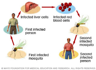
Transmission of malaria, transmission of malaria sporozoites occurs through the bite of a female Anopheles mosquito, the species differ according to geography. Transmission of the asexual forms (trophozoites) causing trophozoite-induced malaria, which can be via blood transfusion (transfusion malaria), via syringe or transmitted from mother to unborn baby melaiui piasen (congenital malaria).

life cycle of malaria

Epidemics of malaria, Malaria is widespread in seiuruh world, either in tropical, temperate and cold climates. To determine the level of malaria endemicity in an area, it must be examined spleen index (spleen index, SI), and the index of parasites (parasite index, PI). In addition it should be investigated to determine the Anopheles mosquito infection rate (infection rate) and the density of mosquitoes (mosquito density). Besides tu socio-cultural life of the population and the environment endemic areas should be studied carefully.

The degree of endemicity of malaria. According to the World Heal Organization (WHO), based on the index override malaria areas are classified into four levels, namely:
Hipoendemis: override index between O to 10 percent;
Mesoendemis: index between li replace up to 50 percent;
Hiperendemis: seialu spleen index above 75 percent with a high index of spleen in adults
Holoendemis: indexes replace always above 75 percent by the spleen index in adults is low. This indicates a strong tolerance of adults against malaria.

Various factors affect the epidemiology of malaria that is the source of infection, either the patient or gametocytes career, namely their capability of transmitting Anopheles mosquito, and sensitive human presence. In endemic areas, especially people with children are the most important source of infection.
Anopheles mosquitoes factors to be considered are the breeding grounds for mosquitoes (breeding places), mosquito longevity and effectiveness in acting as a vector Anopheles transmitters, as well as a dose of sporozoites inoculated each time to suck the blood of patients in both donor and recipient.

The clinical symptoms of malaria, malaria incubation period for each type is different. In malaria vivax and ovale malaria incubation lasts between 10 to 17 days, the falciparum malaria between 8 to 12 days and at malariae malaria, the incubation period lasts between 21 and 40 days.

Malaria show typical symptoms, namely:
Recurrent fever which consists of three stages: freezing (rigor) which lasts between 20 minutes to 1 hour, body heat stage (l-4 hours) and stage sweating a lot (2-3 hours). Splenomegaly and anemia accompanied by malaise.

The pattern of malaria fever. Cycle fever occurs in accordance with the time of skizogeni erythrocytic on each species of Plasmodium.
In tertian malaria, whether malignant or benign, fever takes place every 3rd day (48 hour cycle) and on malariae malaria, fever occurs every 4th day (72 hour cycle). 24-hour cycles of fever can occur if there is a second-generation maturation Pl. vivax within 2 days (called tertian duplex), or third-generation maturation Pl. malariae within 3 days (called kuartana triplex).
A variety of symptoms and complaints of patients can follow the stages of fever, for example, on the stage of rigor, patient shivering despite the patient's body temperature above normal. In stage heat becomes dry skin sufferers, people with red faces and pulse rate increased. Patients also complained of dizziness, nausea, and sometimes vomiting. In children, high fever can cause seizures. At the stadium sweating due to excessive discharge, the patient feels very tired and weak.

Anemia in malaria. Due to rupture of erythrocytes repeatedly during the segmentation process parasites in erythrocytes, patients experience anemia hypochromic microcytic or normocytic hypochromic anemia.
Enlargement of the spleen. One of the important symptoms of malaria are splenomegaly occur after the patient has experienced several bouts of fever. Palpable spleen usually start the second week since the first fever. In the primary malaria, enlarged spleen is unclear and difficult to determine magnifying. The size of an enlarged spleen is important in determining the degree of a malaria endemic area.

Diagnosis of malaria, malaria diagnosis must be determined if found parasites. Examination of peripheral blood is easy to do though sometimes Plasmodium elusive.
Microscopic examination is done with thick drops (thick smear) or with a blood smear (thin-smear). Thick drops performed to determine the rapid diagnosis of malaria, but is undetermined species of the Plasmodium parasite. Blood smear can be used to determine the species of the parasite that causes malaria.

To help with the diagnosis of malaria, especially the concentration of parasites in the blood is very low, performed on peripheral blood serology, for example precipitins test and complement fixation test.
Blood tests also showed a picture of patients decreased hemoglobin, abnormal or decreased leukocytes, platelets decreased, aspartate amino transferase increased, alanine amino transferase increases and increased bilirubin.

Treatment of malaria, anti malaria medication, anti-malaria medicine can be grouped into two groups, namely natural alkaloids, such as quinine and synthetic antimalarial.
Synthetic anti malaria drug is 9-aminoakridin (mepakrin) eg atabrin, kuinakrin, 4-aminokuinoiin (kiorokuin, amodiaquine), 8-aminokuinolin (pamakuin, primaquine), biguanide (proguanil, klopoguanil) and pyrimidine (pyrimethamine). Drug for malaria treatment, Anti malaria drugs can be administered in the form of a combination of pyrimethamine and sulfadoxine are marketed as Fansidar.

Quinine. This natural alkaloid can be administered together with makuin to treat relapsing malaria, acute and severe malaria (given intravenously) or to treat falciparum malaria which is resistant to chloroquine.
Immunity (resistance) to anti malaria drugs. One of the obstacles in combating and controlling malaria is the malaria parasite immunity against anti-malaria drug are used.
A species is declared Plasmodfum still susceptible (sensitive) against certain anti-malarial drugs, if within 7 days of treatment, parasitemi asexual forms had disappeared without a subsequent recurrence (rekrudesensj). This parasite is still sensitive Sensitive stated.
If there has been a parasite resistance to anti-malaria drugs, the malaria drugs should be immediately replaced with other anti-malarial drugs, while malaria control is done by increasing the eradication of malaria vectors are Anopheles mosquitoes.
Prevention of malaria made to the individual and society, namely:
Treat people and people who are sensitive, who resided in endemic areas.
Treating malaria using primaquine career, being able to eradicate gametocytes. However, use of these drugs should not be done en masse because they have side effects.

Preventive treatment in people who will go to malaria-endemic areas
Eradicate the mosquito Anopheles vectors of vector by using appropriate insecticides and destroy the nests of the Anopheles mosquito.
Refrain from mosquito bites by using mosquito nets when sleeping, or using repellen are rubbed on the skin of the body the evening if you're outside the house at night.
Malaria pernicious, pernicious malaria (pernicious malaria) is a collection of symptoms that result from the treatment of falciparum malaria that is not perfect, so the cause of death of patients within one to three days after treatment.
Kanagiutinasi causing any parasite Plasmodium falciparum infected erythrocytes so that the capillaries of various organs unstoppable. This is due to the parasite erythrocytic skizogoni process occurs in the capillary blood vessel organ. The result is a parasitic emboli that are not able to pass through capillaries. Also forms trofozoit and sexual forms of the parasite is attached to each other and also attached to the capillary wall. In pernicious malaria always happen parasitemi weight, either a ring shape or form schizonts.

There are three clinical forms of pernicious malaria, namely cerebral malaria, algid malaria and malaria septikemik.
Cerebral malaria occurs due to brain disorder that causes symptoms of hyperpyrexia, paralysis and coma. In aigid malaria, due to peripheral circulatory failure, patients experience symptoms of collapse with clammy skin. In algid malaria-type gastric collapse accompanied by vomiting, diarrhea koleraik type, and dysentery on the type disenterik.
Septikemik malaria symptoms are the body heat is always high, accompanied by symptoms of pneumonia and cardiac syncope.

Blackwater Fever, Blackwater fever is a form of malaria faiciparum accompanied intravascular hemolysis, fever, and hemoglobinuria.
Blackwater fever is common in patients with malaria faiciparum with the state:
1. Do not have immunity to malaria (non-immune) who received treatment with low doses of quinine.
2. deficiency of glucose-6-phosphate dehydrogenase, so prone to hemolysis of erythrocytes.
In addition, factors low temperatures, fatigue, trauma, pregnant women, childbirth and radiation to the spleen may have an effect on the incidence of blackwater fever.

Pathogenesis Biackwater Fever, a result of the occurrence of intravascular hemolysis, occurs themalbuminemia, hyperbilirubinemia and hemoglobinuria.
Various organ changes, namely:
1. The kidneys become enlarged and dark due to the damming and pigmentation,
2. The liver is enlarged, softened and yellow due to the formation hemosidenin,
3. The gall bladder bile filled with dense dark green,
4. The spleen is enlarged, black for their pigment hemozoin,
5. Hemosidenin many accumulate in the liver, spleen and kidneys.
In times of crisis hemolytic, parasites not found in the blood because it participates destroyed by the process of hemolysis. One week after the crisis ends hemolysis, the parasite can be found in the peripheral blood of patients.

Blackwater Fever diagnosis, blood tests showed normocytic anemia patients with red blood cell count less and 2 million per milliliter, and the hemoglobin is less than 10. During the healing period, it appears the reticulocytosis and lekositosis netrofilik. Biochemical showed increased blood urea, while decreasing cholesterol and greatly decreased haptoglobin.

Complications blackwater fever, blackwater fever Three major complications are renal physiology failure (uremia), liver function failure and circulatory collapse. Blackwater fever death rate of about 20 to 25 percent, mainly due to kidney failure.

Treatment of Blackwater Fever, blackwater fever Every patient should get plenty of rest. Fluid balance must be kept going alkalosis and edema. If needed, brine, parenteral plasma or blood transfusions can be given the appropriate circumstances of the patient. In case of sudden kidney failure, peritoneal dialysis can be given, and in case of hemolytic crisis, give corticosteroids.

Drugs to treat malaria, Anti malaria drug should be administered with caution. Kiorokuin, pyrimethamine or proguanil may be given, but primaquine, kuinakrin, and quinine is contraindicated.
Bacillary Dysentery Disease

1 Comment
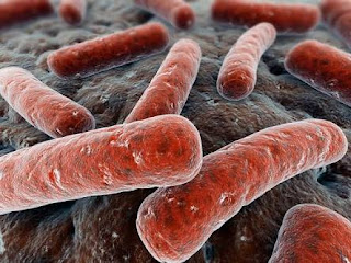
What is the disease Bacillary Dysentery?Bacillary dysentery is an infection of the large intestine by bacteria Shigella, which only cause damage to the intestines and does not cause damage to other organs Network.

bacillary dysentery

Shigella dysenteriae or Shigefia Shigae, is the cause of most violent dysentery for forming endotoxin, often leading to severe epidemics in tropical and subtnopis.

Germs Shigella bacteria are rod-shaped Gram-negative slender, who at coccobacillus shaped bacteria culture bacteria colonies in a 24-hour incubation period, a light translucent colonies are convex, circular with a diameter of about 2 mm.

Sigelosis transmission, transmission of bacillary dysentery occurred because of poor cleanliness and hygiene, their feces patients who become a source of infection, and the presence of flies and insects as vectors transmitting these diseases. Transmission occurs from human patients to others, and the rare transmission of infection from diseased primates to humans.

Clinical symptoms of bacillary dysentery caused by an exotoxin produced by S.dysenteriae of endotoxins produced by other Shigella species. The incubation period lasts between 1 day - 1 week.

Patients with this disease experience sudden high fever accompanied by stomach upsets such as abdominal pain, nausea and vomiting. A few hours later diarrhea that can reach 20-24 times in 24 hours. At first the little stool contains blood and mucus, and then only in the form of blood and mucus,
In severe infection (fulminant type) patients collapse followed by high fever, chills, vomiting, decreased body temperature, severe toxemia, and finally the patient died.

Patients with bacillary dysentery children and elderly people who experienced dehydration and acidosis may also died.
In mild infections, soft stools or normal form, not liquid, blood and mucus, similar symptoms amubiasis.

Sigelosis Diagnosis, Clinical symptoms that occur in different basilen disenteni with clinical symptoms amubiasis. Proctoscopy examination showed diffuse inflammation of the intestinal mucosa, swollen and covered with exudate. Looks shallow ulcers, irregular shapes and sizes covered purulent exudate.

Patient stool culture (and smear the rectum) selective culture medium For example MacConkey or EMB and Thiosulfate-citratbile to follow fermentation test and microscopic examination sigelosis diagnosis.

Serological probes by measuring the increase in antibody titer Shigelfa species can help determine the diagnosis sigelosis.
Treatment and prevention of bacillary dysentery, Sigelosis can be treated with various antibiotics, his eg tetracycline, ampicillin and trimethoprim kioramfenikol sulfametoksasol. Old drug delivery at least 5 days.

Patients should be isolated and disinfected patient excreta. Career sigelosis found should be treated with perfect so do not be a source of disease transmission.

Maintain the cleanliness of food and milk, always cook food and drink, make good excreta disposal systems, combat flies and other insects can prevent the transmission of infectious bacillary dysentery. Supervision of the cleanliness of the food processing industry is mainly done in a residential neighborhood / family must be carried out strictly by preventing the use of raw water to process food and beverages.

Polio: Symptoms, Treatments, Preventions

Add Comment
What's Polio? Polio is also called poliomyelitis or infantile paralysis is a viral infectious disease that attacks the central nervous system and muscles, including the heart muscle. The cause is a virus poliomyelitis virus group which includes entero virus RNA and family Picornaviridae. Capsulated virus has a diameter 20-40Sim virion. There are 3 antigenic types of poliomyelitis virus, types 1, 2 and 3.

polio

The spread of polio, Polio is endemic throughout the world, generally properties without symptoms (asymptomatic) for tenjadinya active immunity in the population. Paralysis that occurs in people with polio is mainly caused by polio virus type 1. The epidemic polio in the past many causes of death of the patient and the occurrence of paralysis due to nerve damage.
Infection occurs through contaminated food or beverage ingredient containing infective polio virus. Human patients are the only source of infection for others, especially careers that are difficult to detect polio which can transmit the virus through direct contact.

Clinical symptoms of polio, After passing through the length of the incubation period is 1-5 weeks, the patient will experience fever, chills, sore throat, severe headache, nausea, vomiting, stiff neck and back pain, followed by paralysis. Paralytic polio can only be diagnosed clinically.

Diagnosis polio, polio To confirm the diagnosis, since AWAI stage disease can be detected viruses cause, namely by breeding stool or throat swab on monkey kidney cell medium. If it happens paralysis, only infective material derived from the digestive tract that still contain the virus.
To help with the diagnosis do serology, among others, for the neutralization test showed increased antibody titers and complement fixation test which showed positive results. Blood tests showed a picture of people with polio mild leukocytosis or normal, whereas the cerebrospinal fluid examination showed high levels of the protein with normal sugar levels.

polio disfunction

Treatment and care polio, polio sufferers, especially children should rest in bed. Symptomatic treatment is given pursuant to patient complaints and clinical symptoms occur.
At the time of the polio epidemic, immediately after the polio virus types cause can be determined, all those who are sensitive to poliodiberi vaccination with the monovalent vaccine Sabin. Giving gammaglobulin hiperimuri at 3-5 weeks before the onset of paralysis can prevent a paralysis. If it happens paralysis, gamma globulin administration hiperimun can not cure paralysis sufferers. In case of severe respiratory disorders, if necessary to do a tracheotomy.

Prevention of polio, vaccination is a very important measure to prevent the spread of polio. Sabin vaccine (live attenuated vaccine) given orally as two doses, the effectiveness of the vaccination can be reached l00%.
Salk vaccine, a formalin-inactivated vaccine given by injection, must be given at least 4 times in a period of several years.
At the time of the polio epidemic, all plans nose and throat surgery had to be postponed. All food waste and excreta polio patients, especially patients with pharyngeal fluid derived from patients should be disposed of properly and covered to avoid spreading the virus by flies and other insects.

Plague Disease: Symtoms and Preventions

Add Comment
What's Plague Disease? Plague disease is a highly contagious infectious disease that often cause epidemics and pandemics are very spacious, with millions of victims died because of it. The cause of the plague is Yersinia pestis bacteria and transmitted mainly by rat fleas (Xenopsylla cheopis).

plague disease bacteria

Yersinia pestis, this plague germs are rod-shaped stocky, with rounded ends. The size is approximately 1.5 x 5.7 microns and coloration are Gram-negative. Germs show pleomorphism properties with an assortment of shapes With staining, visible under the microscope bacteria form bipolar (two poles) that is similar to the pin. Germs do not move, do not form spores and is always covered with slime.
In cultured in medium aerobic or facultative anaerobes, bacteria optimum growth at a temperature of 27 ° C with an optimum pH of 7.2.

Transmission of plague, plague often cause epidemics in many countries, even the plague pandemic that strikes people in dozens of countries around the world, leaving a death toll of millions of lives. Actual bubonic plague is an infectious disease in mice cities (Rattus norvegicus) and mice (Rattus rattus) is the infection spread to humans through the bite of the rat flea (Xenopsylla cheopis and Ceratophyllus fasciatus). In addition plague germs can be transmitted through the air (droplet infection) come and people with pneumonic plague.

Clinical symptoms of Plague
Plague in humans can occur in two forms, namely the shape and form of pulmonary plague bubo plague.


Bubonic plague, After passing through an incubation period of between 2-8 days, groin lymph nodes to swell followed by formation of pus. Patients suffering from fever and cough. If the bacteria enter the bloodstream it will happen septikemi accompanied by bleeding skin and mucous membranes. Sometimes it happens that severe neurological disorder that causes sufferers died.

Pneumonic plague. It also called bubonic plague pneumonia that occurs through the respiratory air transmission causing clinical symptoms such as cough, fever and symptoms of pneumonia accompanied by cough blood. Patients often experience severe cyanosis due to respiratory distress suffered.

Germs can be isolated and Yersinia pestis blood, sputum and pus patients were then examined under a microscope to determine the exact diagnosis of disease plague. To help with the diagnosis do serology plague, for example agglutination test, ELISA, complement fixation test, and immunofluorescent antibody (IFA).

Treatment of plague, In the early stages, patients can be treated with streptomycin, tetracycline or kioramfenikol, with satisfactory results. Without treatment, patients with plague pneumonia will generally die

prevention of Plague, Because it is so contagious, plague sufferers should be isolated and treated immediately. People who had contact with the patient must also be quarantined and closely monitored and given preventive treatment with tetrasikiin or sulfonamides. Everyone who will visit the plague endemic areas should be vaccinated to prevent contracting the disease plague.

Prevention of zoonotic diseases should be accompanied by eradication of rodents and rodent using rodenticides, while the rat fleas eradicated by insecticides.